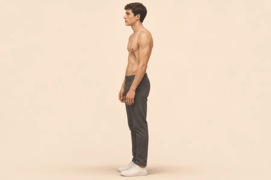
What it is
In pectus excavatum the breastbone (sternum) and the cartilages that join it to the ribs sink inward, leaving a dent in the front of the chest. The dent is usually deepest in the lower part of the sternum and can be symmetric or more marked on one side. It is the most common chest wall deformity: depending on the population studied, 65–95% of cases.
How common it is
Estimates vary a lot, from about 1 in 1,000 to 1 in 250 people. When researchers actually examined people, they found it in 1.3% of schoolchildren aged 11–14 in a Brazilian city, and a CT-based Haller index above 3.25 in 0.5% of adults in a Dallas study.
It is usually reported 3 to 5 times more often in males. The adult CT study found the opposite, so the picture may be less clear-cut than it seems: women are probably under-diagnosed. There is more on this in the guide on pectus in women.
When it appears and how it changes
In a large surgical series the dent was already visible at birth or in the first year in 86% of patients; for many others it is noticed only later. What is consistent is that it often deepens during the growth spurt of puberty. In adulthood it is usually fairly stable. The guide does pectus excavatum get worse with age? covers this in detail.
What causes it
The exact cause isn’t known. The old explanation, that the rib cartilages grow too long and push the sternum back, has been contradicted by 3D CT studies in which the cartilages were the same length as, or shorter than, those of people without pectus. The debate is not closed.
It clearly runs in families: 37% of patients in one series had an affected relative, and up to about half in other reports. Family studies point to several patterns of inheritance, mostly a mix of genes and other factors. About 14 genes have been linked to it, mainly in rare families, and it is part of at least 20 genetic syndromes.
Related conditions
- Scoliosis: about one in four to five people with pectus excavatum in surgical series.
- Marfan syndrome: about 1–5% of people with a pectus deformity, more in mixed forms. The 2024 UK guideline estimates that about 5% have a connective tissue disorder and recommends checking eyes, heart, skeleton and family history, with referral when in doubt.
- Ehlers-Danlos and Noonan syndromes are associated too, but there is no reliable figure for how often.
- Poland syndrome (a missing chest muscle on one side) is rare: 3 in 704 patients in one series.
Features such as long limbs, very flexible joints, a high-arched palate or flat feet, especially with a family history of heart or aortic problems, are worth mentioning to a doctor.
Symptoms
Many people have no physical symptoms. When they do, they usually appear during exercise rather than at rest:
- less stamina than friends, getting out of breath sooner;
- chest pain;
- palpitations or a racing heart;
- tiredness.
The effect on how people feel about their body is often the heaviest part. Compared with people without pectus, patients report a more negative body image and a lower physical quality of life, without more psychiatric disorders. The UK guideline recommends asking about it as part of the assessment.
The heart
The sunken sternum can press on the right side of the heart, which then fills less well and can’t raise its output as much during exercise. In 168 adults, an ultrasound during surgery showed the right heart chambers widening and right-sided output rising by 38% once the chest was corrected.
Whether this changes fitness is less clear: some studies found a better maximum oxygen uptake after surgery (8–15% higher), others found no change, and a meta-analysis found an overall benefit. In a small study of teenage boys, fitness was lower than in peers on average, with a lot of overlap. Mitral valve prolapse (a floppy heart valve) and rhythm disturbances are reported in some surgical series; serious arrhythmias only in extreme cases.
The lungs
Lung function is usually normal or slightly reduced. In 310 patients, about 1 in 7 had a mild restrictive pattern (the lungs hold a little less air), and the risk rose with severity. After surgery, vital capacity improved modestly in one large study and didn’t change significantly in an earlier meta-analysis.
How it’s diagnosed
- Examination: a doctor looks at the shape, its depth and symmetry, posture and signs of related conditions.
- CT or MRI: recommended by the UK guideline when treatment is being considered, ideally taken after breathing out, because the index can be up to 30% higher after breathing in. From the images come the Haller index (above 3.25 is the classic threshold for a severe form) and the correction index.
- Chest X-ray: can give a similar index, but the evidence comes from a small study.
- Heart ultrasound and lung function tests to rule out other causes of breathlessness, and a cardiopulmonary exercise test in severe forms with exercise intolerance. The guideline also suggests an ECG.
For a first idea of where you stand, the self-check takes one minute.
Is it dangerous?
Most cases are mild, and effects on the heart and lungs, when present, usually show only during hard exercise. Very rarely, extreme forms cause dangerous heart rhythms, fainting or near-fainting, compression of the large vein that returns blood to the heart, or difficulty swallowing. See a doctor promptly for chest pain or fainting during exercise, palpitations, or breathlessness out of proportion to effort.
Does it go away?
There is no evidence that it disappears on its own. Many mild forms, however, need no treatment beyond keeping an eye on them, especially in adulthood.
Treatment, in brief
- Observation is enough for most people.
- Exercise and physiotherapy improve posture, mobility and strength; there is no solid evidence that they change the shape of the bone. The exercise program explains what is known.
- The vacuum bell is safe; in two series about 1 in 5 reached an excellent correction and about a third an excellent or good one, with better results in children under 11, in shallower dents and with more than a year of use. See the vacuum bell guide.
- Surgery (Nuss or Ravitch) gives the most solid correction for marked forms: the surgery guide explains when it’s considered.